Anatomy of the eye
The Structure of the Eye
The eye is made up of three layers:
-
The fibrous tunic, which consists of the sclera and the cornea.
-
The vascular tunic which consists of the iris, the choroid and the ciliary body and is
responsible for nourishment.
-
The nervous tunic which is the inner layer of photoreceptors and neurons which consists of
the retina.
The nervous tunic or neural layer contains the special photo (light) receptors known as rods and cones.
-
Rods do not discriminate between different colors of light. The rods let us see at twilight and in dimly lit rooms.
-
Cones do discriminate colors. Cones require brighter light to function than rods do.
There are approximately 120 million rods spread towards the outside of the retina and about 6 million cones concentrated near the center of the macula. The highest concentration of cones is found at the fovea which is the center of the macula. The macula is where light will focus in a healthy eye.
The point where the optic nerve enters the eye is known as the optic disc. The optic disc does not contain receptor cells so it is sometimes called the "blind spot".
The eye also contains three fluid-filled chambers:
-
The anterior chamber between the cornea and the iris
-
The posterior chamber between the iris and the lens
The anterior and posterior chambers contain a fluid called aqueous humor. Aqueous humor is watery fluid produced by the ciliary body. It maintains pressure (called intraocular pressure or IOP) and provides nutrients to the lens and cornea. Aqueous humor is continually drained from the eye through the Canal of Schlemm.
3. The vitreous chamber is found between the retina and the lens and is filled with a thicker gel-like substance called vitreous humor which maintains the shape of the eye.

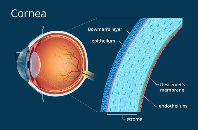
Light enters the eye through the transparent, dome shaped cornea.
The cornea consists of five distinct layers:
-
The outer most layer is the epithelium which rests on Bowman’s membrane
-
The next layer, which acts as a protective barrier, is Bowman's Membrane
-
The stroma which is between the two membranes makes up 90% of the thickness of the
cornea
-
Descemet's Membrane separates the stroma and the endothelium.
-
The inner most layer, the endothelium, removes water from cornea, helping to keep the
cornea clear.
Memory Hint: B comes before D and think “end” for endothelium.
From the cornea, light passes through the pupil. The amount of light allowed through the pupil is controlled by the iris, the colored part of the eye.
The iris has two muscles:
-
The dilator muscle which opens the iris allowing more light in
-
The sphincter muscle which closes the iris
The iris has the ability to change the pupil size from 2 millimeters to 8 millimeters.
Just behind the pupil is the crystalline lens. The purpose of the lens is to focus light on the retina. The process of focusing on objects based on their distance is called accommodation. The closer an object is to the eye the more power is required of the crystalline lens to focus the image on the retina. The lens achieves accommodation with the help of the ciliary body which surrounds the lens. The ciliary body is attached to lens via fibrous strands called zonules.
When the ciliary body contracts, the zonules relax allowing the lens to thicken, adding power, allowing the eye to focus up close. When ciliary body relaxes, the zonules contract, drawing the lens outward, making the lens thinner, and allowing the eye to focus at distance.